The majority of limb bones develop through a process known as endochondral ossification, which begins with the formation of an undifferentiated cartilage model, called chondrocytes. These chondrocytes go through stages of differentiation into resting zone (RZ), proliferative zone (PZ), and hypertrophic zone (HZ) chondrocytes, resulting in a well-structured growth plate. This organized growth plate is essential as it aligns along the longitudinal axis of the future bone, serving to preconfigure the initial shape of each future bone.
A significant reduction in the height of the human ilium compared to apes has prompted research into its chondrification during crucial developmental stages, specifically between gestational days 45 and 72 (E45–E72). At E45, the ilium is primarily composed of undifferentiated chondrocytes that are longitudinally aligned. By E53–E54, rapid proliferation and differentiation of these chondrocytes occur, shifting the growth plate alignment to a horizontal (transverse) orientation by E72. This growth pattern reveals a unique mechanism whereby the ilium's growth plates expand perpendicular to its original bone axis, contributing to the ilium's width while reducing its height.
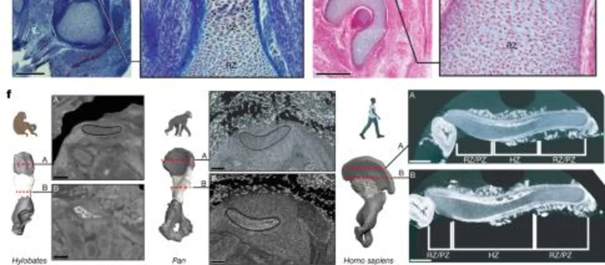
To understand the evolutionary aspects of the human ilium's morphogenesis, comparisons were made with the iliac growth plates of laboratory mice and other primates. Histological examinations showed that mouse, mouse lemur, and tamarin monkey ilia have RZ, PZ, and HZ oriented longitudinally. In contrast, the human ilium's growth plate orientation has shifted towards a transverse alignment. This shift is particularly notable as it differentiates human growth plate development from that of other primates.
To analyze the molecular basis of these developmental changes, single-cell multiomics and spatial transcriptomics were utilized on micro-dissected ilia and adjacent soft tissues from E45–E72. The analyses revealed a diverse array of cell types including mesodermal cells, chondroprogenitors, and osteoblasts, with significant changes in cellular composition over time. Key genes such as SOX9 and its effector PTH1R were identified as critical players in chondrification, with SOX9 showing a spatially asymmetric expression pattern during key developmental stages.
In patients with campomelic dysplasia caused by SOX9 mutations, ilia displayed notable narrowing, supporting the hypothesis that SOX9 and PTHrP signaling are integral to ilium morphology. Similarly, mutations in PTH1R have been associated with various skeletal disorders, leading to insights about the genetic underpinnings of human iliac development and its distinctive transverse growth pattern.
In addition to intrinsic molecular cues, external signaling mechanisms were examined, particularly the interactions between iliac chondrocytes and adjacent mesenchymal tissues. Pathways such as pleiotrophin (PTN) and midkine (MK) were identified, both of which are involved in chondrocyte migration and proliferation. A further pathway involving parathyroid hormone (PTH) was discovered to signal from the perichondrium to iliac chondrocytes, indicating a complex network of interactions that drive the ilium's anterior-posterior expansion.
The research also highlights the evolutionary signals that govern the shift in growth plate orientation in the human ilium. Through the integration of single-cell sequencing data with evolutionary genomic analyses, strong overlaps were identified between human accelerated regions (HARs) and regulatory elements crucial for chondrocyte differentiation and skeletal morphogenesis.
Human ossification patterns differ significantly from those of other primates. For instance, the primary ossification center (POC) in the human ilium initiates in a unilateral manner compared to the bilateral ossification seen in other mammals. This unique pattern of ossification is characterized by a delay in the internalization of bone formation, which occurs radially along the periphery of the cartilage, contrasting with the typical internal ossification processes observed in long bones.
Overall, the findings elucidate a complex interplay of genetic, cellular, and evolutionary dynamics that underlie the unique development of the human ilium. The integration of multiomic analyses provides a comprehensive view of the gene regulatory networks that influence the skeletal morphology, offering valuable insights into how evolutionary changes can impact developmental processes.