Over the past seven years, a global collaboration of more than 150 scientists has undertaken the most complex neuroscience experiment to date, culminating in the release of their findings this week. The MICrONS Project—short for Machine Intelligence from Cortical Networks—has successfully completed the first phase of an ambitious goal: to construct a functional wiring diagram of a portion of the brain. This monumental achievement has been published in the journal Nature, featuring a comprehensive collection of ten groundbreaking studies.
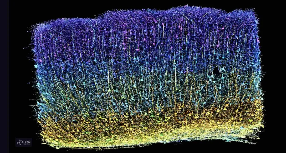
The resulting 3D wiring diagram is vast, encompassing a staggering 1.6 petabytes of data—equivalent to 22 years of continuous HD video. This data provides an unparalleled glimpse into brain function and the intricate organization of the visual system. The research initiated at Baylor College of Medicine, where scientists utilized specialized microscopes to monitor brain activity from a one cubic millimeter portion of a mouse’s visual cortex while the subject viewed various movies and YouTube clips. Following this, researchers from the Allen Institute meticulously sliced the same cubic millimeter of brain tissue into over 25,000 sections, each measuring 1/400th the width of a human hair, and employed high-resolution electron microscopy to capture detailed images.
The MICrONS Project has produced the most detailed wiring diagram of a mammalian brain to date, which is now freely accessible online. David Markowitz, Ph.D., who led the project after his tenure at the IARPA (Intelligence Advanced Research Projects Activity), which partially funded the initiative, described this achievement as “a watershed moment for neuroscience, comparable to the Human Genome Project.” Meanwhile, another team at Princeton University harnessed artificial intelligence and machine learning to reconstruct the cells and their connections into a detailed 3D volume. This reconstruction includes an astonishing 523 million synapses (the connection points between 200,000 cells) and spans four kilometers of axons (the branches that extend to other cells).
Clay Reid, Ph.D., a senior investigator and pioneer in the field of electron microscopy connectomics, emphasized the significance of these findings, stating, “Inside that tiny speck is an entire architecture like an exquisite forest.” This research unveils various rules of connections that align with existing neuroscience theories and opens avenues to test these theories while potentially uncovering new insights.
The studies have revealed new cell types, characteristics, organizational structures, and functional principles, including an unexpected new principle of inhibition within the brain. Contrary to previous beliefs that categorized inhibitory cells as a simplistic dampening force, researchers discovered a more intricate system of communication. Inhibitory cells exhibit selectivity in their actions; they coordinate with one another to suppress multiple excitatory cells while targeting specific types of cells.
Understanding the brain's structure and function, along with analyzing the connections between neurons at such an unprecedented scale, opens new possibilities for research into brain disorders such as Alzheimer’s, Parkinson’s, autism, and schizophrenia, which are characterized by disrupted neural communication. Nuno da Costa, Ph.D., an associate investigator at the Allen Institute, remarked, “If you have a broken radio and you have the circuit diagram, you’ll be in a better position to fix it.” This research serves as a “Google map” or blueprint of a grain of sand-sized portion of the brain, enabling comparisons of healthy brain wiring against models of diseases.
This extensive collaboration, which also included scientists from Harvard University, was made possible through support from the IARPA and the US National Institutes of Health’s BRAIN Initiative. Markowitz emphasized that these foundational scientific insights into brain wiring are crucial for advancing our understanding of brain injuries and diseases, ultimately paving the way for new treatments and cures.
In a notable contrast to Francis Crick's assertion in 1979 that creating an exact wiring diagram for a cubic millimeter of brain tissue would be impossible, the MICrONS Project has made significant strides towards this once-unimaginable goal. Clay Reid, who dedicated his career to this pursuit, expressed optimism that this detailed map of neuronal connectivity, form, and function is a crucial step toward unraveling the origins of thought, emotion, and consciousness.
The MICrONS Project not only exemplifies a remarkable scientific breakthrough but also brings us closer to understanding one of the most complex systems known to humanity—the brain.