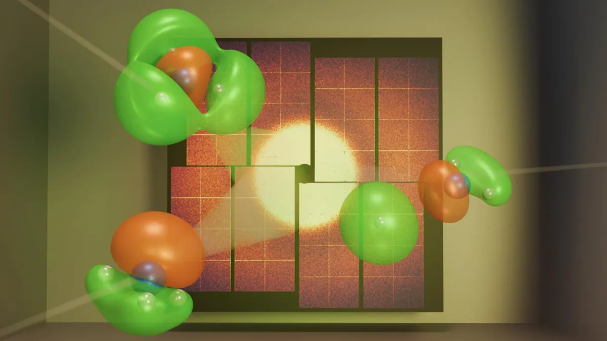
In a groundbreaking study published on August 20 in the journal Physical Review Letters, scientists have successfully utilized ultrafast X-ray flashes to capture a direct image of a single electron as it moved during a chemical reaction. This remarkable achievement marks a significant milestone in the field of quantum physics and provides invaluable insights into the dynamics of chemical reactions.
The researchers focused on the behavior of a valence electron, which is located in the outer shell of an atom. This study specifically examined how a valence electron moved when an ammonia molecule broke apart. Traditionally, scientists have employed ultrafast X-ray scattering techniques to image atoms and their reactions, using incredibly short bursts of X-rays to freeze fast-moving molecules in action. X-rays are favored for their ideal wavelength range, which is perfect for capturing intricate details at the atomic scale.
However, a challenge arose as X-rays predominantly interact with core electrons, which are situated near an atom’s nucleus. As a result, the valence electrons—those responsible for driving chemical reactions—remained obscured. Ian Gabalski, a physics doctoral student and the lead author of the study, emphasized the importance of capturing the actual electrons responsible for these motions. He stated, “If scientists can understand how valence electrons move during chemical reactions, it could help them design better drugs, cleaner chemical processes, and more efficient materials.”
To embark on their investigation, the research team needed to identify a suitable molecule for imaging. They settled on ammonia, which Gabalski described as "kind of special." Due to its composition of primarily light atoms, there were fewer core electrons that could obscure the signals from the outer valence electrons. This unique property provided the team with a significant opportunity to visualize the movement of the valence electron.
The experiment took place at the SLAC National Accelerator Laboratory's Linac Coherent Light Source, a facility known for producing intense, short X-ray pulses. The process began by exposing the ammonia molecule to a brief burst of ultraviolet light, which elevated one of the electrons to a higher energy level. In molecules, electrons typically reside in low-energy states, and pushing them to a higher state initiates a chemical reaction.
Following this excitation, the researchers employed X-ray beams to document how the electron cloud evolved as the ammonia molecule began to disintegrate. Gabalski explained that, in quantum physics, electrons do not behave like tiny particles orbiting the nucleus; rather, they exist as probability clouds. The density of these clouds indicates the likelihood of locating the electron, giving rise to the concept of orbitals—distinct shapes that define the energy and position of electrons within a molecule.
To accurately map this electron cloud, the team conducted quantum mechanical simulations to determine the electronic structure of the ammonia molecule. Gabalski elaborated, “This program figures out where the electrons are filling up those orbitals around the molecule.” When the X-rays traverse the electron's probability cloud, they scatter in various directions, creating an interference pattern that the team could analyze.
By measuring this interference, the researchers successfully reconstructed an image of the electron's orbital, revealing its movement throughout the chemical reaction. They compared their findings against two theoretical models: one that factored in valence electron motion and another that did not. The results aligned with the first model, confirming that they had indeed captured the electron's rearrangement in real-time.
Looking ahead, the researchers aspire to adapt their imaging system for use in more intricate, three-dimensional environments that better replicate real biological tissues. This advancement could pave the way for innovative applications in regenerative medicine, such as the ability to grow or repair tissue on demand, highlighting the potential of this groundbreaking research in transforming the fields of chemistry and medicine.