In a significant advancement in the field of neuroscience, scientists have unveiled the most detailed maps of how our brains differentiate from stem cells during the crucial stages of embryonic development and early life. This groundbreaking research, compiled in a series of five papers published in the prestigious journal Nature on November 5, highlights the tracking of hundreds of thousands of early brain cells in the cortexes of both humans and mice. The researchers captured, with unprecedented precision, the molecular events that lead to the formation of a diverse mixture of neurons and supporting cells.
“It's really the initial first draft of any ‘cell atlases’ for the developing brain,” stated Hongkui Zeng, the executive vice-president and director of the Allen Institute for Brain Science in Seattle, Washington, and a co-author of two papers in the collection. This innovative approach marks a pivotal moment in our understanding of brain development and its implications for neurological health.
These newly created atlases provide promising avenues for studying various neurological conditions such as autism and schizophrenia. With this wealth of data, researchers can now “mine the data, find genes that may be critical for a particular event in a particular cell type and at a particular time point,” according to Zeng. “We have a very exciting time coming,” adds Zoltán Molnár, a developmental neuroscientist at the University of Oxford, UK, who was not involved in the studies.
This pioneering work is part of the BRAIN Initiative Cell Atlas Network (BICAN), a project launched in 2022 by the Brain Research through Advancing Innovative Neurotechnologies (BRAIN) Initiative at the US National Institutes of Health. The initiative has received US$500 million in funding to construct reference maps of mammalian brains, enhancing our understanding of brain structure and function.
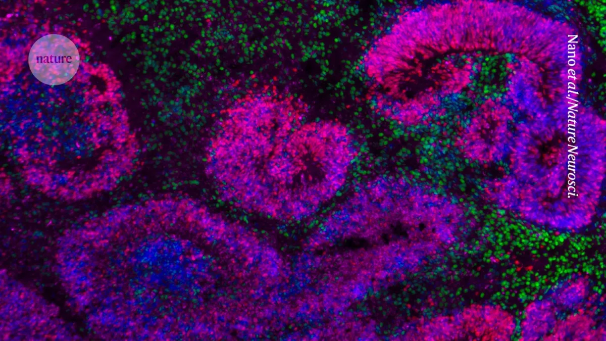
Two of the papers focus specifically on mapping parts of the mouse’s cerebral cortex, the area of the brain responsible for cognitive functions and perception. Zeng and her colleagues meticulously documented the development of the visual cortex from 11.5-day-old embryos to 56 days after birth. They successfully created an atlas comprising 568,654 individual cells, identifying 148 cell clusters and 714 subtypes. “It's the first complete high-resolution atlas of cortical development, including both prenatal and postnatal phases,” Zeng asserted.
One unexpected finding from their research was the extent to which neurons continued to adopt specialized identities after birth, particularly during the critical period from day 11, when newborn mice open their eyes for the first time, to day 21.
Another study concentrated on brain development in human embryos, addressing the long-standing question of how different brain cells emerge. “How different brain cells emerge is a long-standing question in developmental neurobiology,” explained Tomasz Nowakowski, a neuroscientist at the University of California, San Francisco. His team created a family tree map of human cortical cells using brain tissue collected from eight human fetuses at various stages of development.
Through innovative methods, they identified 6,402 progenitor cells within these tissues and introduced a unique DNA ‘barcode’ into each cell. This technique allowed researchers to track which stem cells generated specific types of brain cells, as the barcodes are replicated and passed on during cell division. Nowakowski’s team discovered that in embryos younger than 20 weeks, progenitor cells primarily developed into excitatory neurons, responsible for relaying electrical signals to activate other neurons. However, after 20 weeks, there was a notable shift toward generating inhibitory neurons, which serve to halt neuronal communication. “This shows how these progenitors transition from one type into the other,” Molnár noted.
The team also observed that the generation of other brain cell types—specifically astrocytes and oligodendrocytes—in humans appears to be “much more continuous and protracted” compared to that in mice, as Nowakowski highlighted. “We actually don't know what controls all these developmental programs and switches or why they occur in development and especially at such precise moments,” he added. Addressing these fundamental questions will be a key goal for future studies, paving the way for deeper insights into brain development and its implications for human health.