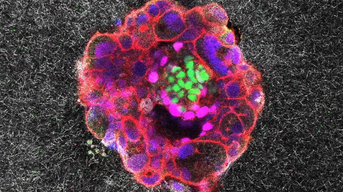
A groundbreaking time-lapse film has provided unprecedented insight into a critical milestone of human development: the moment when a newly formed embryo latches onto the uterine lining. In a significant breakthrough, researchers have successfully captured real-time footage of an embryo engaging with a high-fidelity replica of the uterine lining, illustrating how it burrows itself into its new home and remodels the environment around it. The findings of this important study are featured in the latest issue of Science Advances.
The authors of the study were motivated to simulate the implantation process due to the challenges involved in capturing these crucial events in real life. Co-author Samuel Ojosnegros, a bioengineer at the Institute for Bioengineering of Catalonia (IBEC) in Barcelona, Spain, emphasized the difficulty of studying implantation, stating, “It’s very inaccessible because it’s all happening inside the mother. It’s such an important process for human reproduction, but at the same time, we don’t have the technology to study it.”
Previous research has explored how human embryos interact with glass surfaces, but such materials do not allow embryos to penetrate as they would with real human tissue. To overcome this limitation, the research team devised a more lifelike mock-up by creating a faux uterine lining made from a gel abundant in collagen and proteins essential for embryonic development. This innovative approach was key to accurately observing the complex interactions during implantation.
To produce their stop-motion film, the researchers placed human embryos donated by a local hospital in proximity to the gel. As the embryo attached to the faux uterine lining, the team meticulously captured images approximately every 20 minutes over the course of 16 to 24 hours. These still images were then stitched together to create the time-lapse footage that offers a rare glimpse into the implantation process.
Co-author Amélie Godeau, a biomechanics researcher at IBEC, expressed her astonishment at the speed with which the embryo embedded itself into the gel. “My first reflex was to think my experiment had gone wrong and there was some drift in the microscope,” Godeau recounted, highlighting the unexpected dynamics of the implantation process. In contrast, the team observed that mouse embryos tend to adhere to the surface of the uterus rather than embedding themselves within it, underscoring the unique aspects of human embryonic development.
This innovative research not only enhances our understanding of the human implantation process but also opens new avenues for studying reproductive health and embryonic development. By utilizing advanced techniques and creating a realistic environment, the researchers have paved the way for future studies that could lead to improvements in reproductive technology and therapeutic interventions.